Imaging
Technology You Can Count On
Howard Memorial Hospital knows it’s critical to have access to top-of-the-line diagnostic technology when medical imaging needs arise. We’re committed to providing expert diagnostics, ensuring a high level of insight and care for all our patients.
Currently, HMH is one of only a few hospitals in Arkansas that offer digital imaging for a wide range of diagnostic procedures. This gives us an advantage in providing physicians with clean, clear images, so they can quickly determine the extent of an injury or medical condition.
Imaging Services We Provide
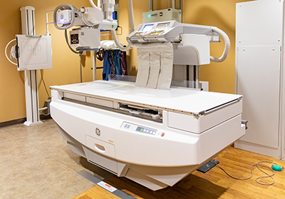
Radiology (x-ray) is a technique that generates and records an x-ray pattern, so a static image is created after exposure to the x-ray. During this procedure, an x-ray beam is passed through the body, and a portion of the x-ray is absorbed or scattered by the body’s internal structure. The remaining x-ray pattern is transmitted to a detector, so an image can be recorded for later evaluation.
This recording shows images of the body’s internal structure, so our providers can assess the presence or absence of disease, foreign objects, structural damage or an anomaly. This allows us to better diagnose and treat patients.
Also known as dual-energy x-ray absorptiometry (DEXA), this is a form of imaging that uses tiny amounts of ionizing radiation to measure bone mineral density, so we can determine an individual’s risk for bone fractures or determine if there is an osteoporosis diagnosis.
The radiation amount used in this bone-density test is less than one-tenth of a more traditional chest x-ray and less than a day’s worth of natural radiation exposure.
This is a type of medical imaging that shows a continuous x-ray image on a monitor. Fluoroscopy is used by our providers to diagnose or treat patients by displaying the specific movement of a body part, a type of x-ray contrast, through the body.
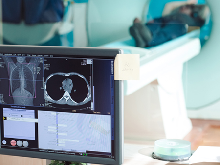
Computed tomography scanning, also known as CT scanning, is a medical imaging procedure that uses x-rays to show cross-sectional images of the body, producing images in a way to help deliver useful information. During a CT scan, the patient undergoes several consecutive and simultaneous x-rays that can be configured as a three-dimensional reconstruction of the body part being imaged.
A CT scan does deliver more ionizing radiation to the patient than a simple x-ray, but this ensures CT scans can better distinguish between different types of body tissues than a normal x-ray. Given CT scanning’s ability to image large areas over a short period of time, it offers much better resolution of many different body structures in a range of different spatial configurations. Many times, a CT scan is performed using x-ray dye or another contrast agent, which is administered by mouth or by vein. This helps our providers identify the intestines or vasculature and assists in the diagnosis of disease or injury.
Ultrasound imaging (also known as sonography) uses high-frequency sound waves to help our providers view soft tissues like muscles and internal organs. Ultrasound images are captured in real-time, showing movement of the body’s internal organs and blood flowing through blood vessels. While there is no documented evidence of ultrasound dangers to either patients or staff, HMH is cautious about the frequency of use, especially with pregnant patients.
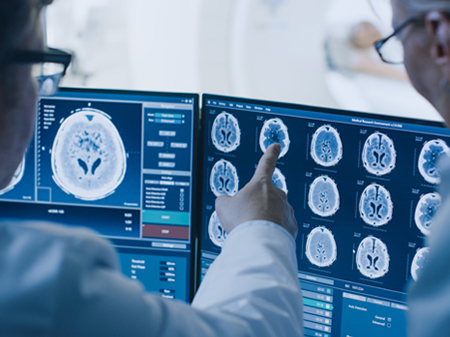
Magnetic resonance imaging (MRI) is a medical imaging procedure that uses strong magnetic fields and radio waves to produce cross-sectional images of organs and internal structures of the body. The signal detected by an MRI machine varies depending on the water content and local magnetic properties of a specific area of the body. This helps our providers in distinguishing different tissues or substances from one another when studying the image.
MRIs give different information about structures of the body than an x-ray, ultrasound or CT scan. With most MRIs, an electric current is passed through coiled wires to create a temporary magnetic field in the patient’s body. Radio waves are then sent from, and received by, a transmitter/receiver in the machine, and these signals are used to produce digital images of the specific body area of interest.
Nuclear medicine uses radioactive material to diagnose and treat a wide variety of diseases and conditions. With a diagnostic nuclear medicine study, a patient inhales, swallows or receives an injection of a small amount of a radiopharmaceutical (a drug with a radioactive component) that accumulates in a specific organ or area of the body.
The energy that is emitted by the radioactive material is detected by a device, then processed and measured by a computer. After that, this energy is displayed as an image on a screen, which one of our providers interprets. An image like this provides information on the structure and function of specific organs and tissues.
Positron emission tomography-computed tomography (PET/CT) combines nuclear medicine techniques with CT technology. The PET/CT machine delivers, detects and processes multiples images at once and combines various imaging modalities (hybrid imaging).
Hybrid imaging provides HMH with detailed information and accurate diagnoses. PET/CT is particularly useful in the diagnosis and treatment of cancer.
Using low-energy X-rays, mammography is the process of examining the human breast for diagnosis and screening with the goal of detecting any possible signs of breast cancer, such as masses or microcalcifications. Mammograms utilize doses of ionizing radiation to create images, which are then reviewed and analyzed for anything abnormal.
At age 40, women should start getting mammograms every year. At a much earlier age, women should talk to their doctors about potential risk factors they may have, such as family members with breast cancer, genetic mutations, previous radiation treatment for cancer and more. If a patient is at high risk due to these factors, a doctor may recommend starting breast cancer screenings as early as age 25.
If you have any questions, comments or concerns regarding our diagnostic imaging department, please contact our radiology director: Stacy Harberson at 870-845-8076.
Your Medical Imaging and Records
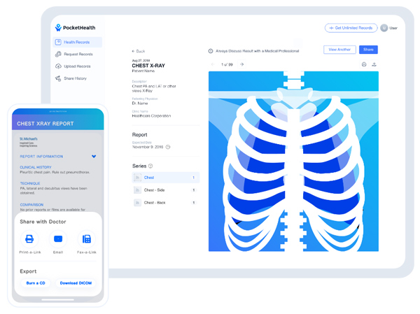
Create an account through PocketHealth and have access to your medical images, share them with your doctors and get involved in your own care.
Please note, you will need your Medical Record Number to sign up for an account with PocketHealth.
With an account, you’re able to:
• Access your images from any device in full diagnostic quality.
• Share images easily with any of your doctors.
• Ensure doctors can make fast diagnoses, directly in the PocketHealth system.
• Store images permanently, so you can view them again at any time.
• Own your own healthcare journey by staying informed about your diagnosis and hearing from doctors directly.
Create a secure account through PocketHealth here.
Mobile MRI
We have a mobile MRI service, available Monday through Friday, at HMH. Your physician can easily schedule an MRI appointment for you through this mobile service by calling our central scheduling department at 870-845-8156.
Mobile PET/CT
We have a mobile PET/CT service, available on the first Saturday of each month, at HMH. Your physician can easily schedule a PET/CT appointment for you through this mobile service by calling our central scheduling department at 870-845-8156.